.webp)
A brain MRI scan is a non-invasive imaging procedure that uses a powerful magnet and radio waves to create highly detailed pictures of your brain. It’s one of the most effective tools for diagnosing a wide range of neurological conditions without using any radiation.

Think of trying to understand how a complex engine works without ever being able to look inside. A brain MRI scan gives doctors that essential window, offering an incredibly clear view of the brain’s soft tissues—something X-rays and CT scans just can't capture with the same detail.
It’s like creating a high-resolution, slice-by-slice map of your brain. This allows specialists to examine its structure with remarkable precision, from the tiniest nerves to the major blood vessels. The technology itself is fascinating; it uses a powerful magnetic field to neatly align the protons in your body’s water molecules. Radio waves then briefly knock them out of alignment, and as they snap back into place, they send out signals. A computer translates these signals into the detailed images we see.
It’s not every day a doctor suggests a brain MRI. They’re typically recommended to investigate symptoms that don’t have an obvious cause, making them a crucial tool for both diagnosis and monitoring.
Exploring your symptoms with a specialist is the best way to know if an MRI is the right next step. You can get a better sense of how experts approach these conditions by understanding the field of neurology.
An MRI can help get to the bottom of persistent or puzzling symptoms. The table below outlines some of the most common reasons a specialist might refer you for a scan.
This isn’t an exhaustive list, but it highlights how an MRI helps connect symptoms to a concrete diagnosis, paving the way for effective treatment.
A brain MRI provides an unmatched view of the brain’s soft tissues, making it the gold standard for detecting subtle changes that could signal serious conditions. It helps turn diagnostic uncertainty into a clear plan for treatment.
Here in the UK, the brain MRI is a cornerstone of modern diagnostics, but it remains a specialised procedure. Just to give you an idea, in April 2025, NHS England recorded 380,000 MRI scans of all types. In that same month, they performed 1.84 million X-rays.
This difference shows that while MRIs are essential, they are used more selectively. Their complexity and cost mean they are reserved for cases where those incredibly detailed images can provide the most crucial health insights, guiding specialists to the right diagnosis and treatment. You can read more about these diagnostic imaging statistics on the NHS England website.
Knowing what to expect before your brain MRI scan can make all the difference. A little preparation turns a potentially daunting experience into a smooth, straightforward process, helping you walk into your appointment feeling calm and confident.
For most scans, you won't need to do anything special. You can usually eat, drink, and take your regular medications as normal. If your particular scan requires a different approach, the medical team will give you very clear instructions. Just be sure to follow their guidance to the letter—it ensures they get the best possible images.
This is probably the most important part of your preparation. The MRI machine is built around an incredibly powerful magnet, which means no metal is allowed anywhere near it. The easiest thing to do is leave all your jewellery, watches, and hairpins at home.
When it comes to clothes, think comfort and simplicity. Loose-fitting clothing without any metal parts is ideal. That means steering clear of:
Leggings, tracksuits, or simple cotton tops are all great choices. If you arrive already wearing metal-free clothes, you often won't need to change into a hospital gown, making things quicker and more comfortable for you. For more specific advice on what to expect during your visit, you can find extra patient information here.
Your safety is the absolute top priority. Because the powerful magnetic field can interact with any metal inside your body, it’s vital to tell the radiography team about any medical implants or devices you have before the scan begins.
You'll be asked to fill out a detailed safety questionnaire before your appointment. It is absolutely essential to answer every question accurately, even if you think something isn’t relevant. This is the single most important step in ensuring your scan is safe.
You must let the team know if you have any of the following:
Giving the team this information allows them to take the right precautions and confirm it’s safe for you to have a brain mri scan. And if you're feeling anxious or claustrophobic, please tell them. They're brilliant at helping people feel at ease and can offer support, like playing music through your headphones or giving you regular updates through the intercom while the scan is running.
Knowing what happens step-by-step during a brain MRI scan can make the whole experience feel much less daunting. It’s a lot easier when you understand what’s going on, from the moment you walk into the scanning room to the final click of the machine.
A radiographer will help you get comfortable on a motorised bed, which then gently glides into the centre of the large, tunnel-shaped scanner. Don't worry – the opening is well-lit and ventilated. While it can feel a bit snug, it’s all designed for safety and to get the clearest pictures possible. Your main job is simply to lie as still as you can, because even tiny movements can blur the images.
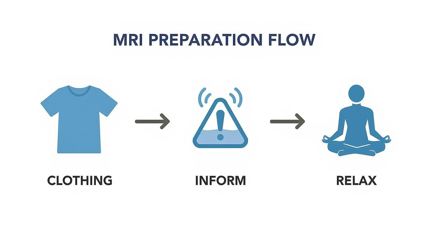
The infographic below gives you a quick visual rundown of the simple but important prep steps right before your scan.

As you can see, it really comes down to a few key things: wearing metal-free clothing, double-checking your medical history with the staff, and just taking a moment to relax. Getting these right helps ensure everything goes smoothly.
Once you’re settled, the radiographer will place a special piece of equipment called a head coil around your head. It might look a bit like a helmet, but it doesn't actually touch you. Think of it as an antenna, designed to capture the highest quality signals from your brain.
You’ll also get headphones or earplugs to block out the noise. An MRI machine is incredibly loud, making a series of strong clanking, buzzing, and whirring sounds while it works. This is completely normal; it’s just the sound of powerful magnets switching on and off to create the images.
The radiographer will be in the next room, watching you through a large window and talking to you over an intercom. They'll keep you updated the whole time, letting you know how long each sequence of scans will last. You'll also be given a buzzer or panic button to hold, so you can get their attention immediately if you need anything.
The whole thing usually takes between 30 and 60 minutes. This is a bit longer than other types of scans. If you’re wondering how that stacks up, you can read our guide on how long a CT scan takes.
Sometimes, your doctor might ask for a contrast agent to be used during the scan. This is a special dye, usually containing a substance called gadolinium, which is given through a small IV line in your arm.
A contrast agent helps to highlight specific areas within the brain, making blood vessels, tumours, or areas of inflammation appear much brighter and clearer on the final images. This added detail can be vital for an accurate diagnosis.
If you need a contrast dye, it’s usually administered part-way through the scan. It’s a very common and safe part of many MRI procedures. You might feel a cool sensation in your arm as it goes in, but this passes in a moment.
Once the scan is finished, the radiographer will slide you out of the machine, and you’ll be free to get on with your day.
It’s completely normal to have questions about safety when you’re facing any kind of medical procedure. When it comes to a brain MRI scan, the most important thing to know is that it’s an exceptionally safe imaging technique, trusted by doctors all over the world for its accuracy and non-invasive approach.
The main reason it’s so safe is that MRI technology doesn't use any ionising radiation. Unlike X-rays or CT scans, which use small, controlled doses of radiation to see inside the body, an MRI relies on a powerful magnetic field and radio waves. This completely removes the risk of radiation exposure, making it a safe option for repeated use and even for children or pregnant women (after the first trimester).
The biggest safety factor to consider is the scanner's incredibly strong magnet. Just think of it as a super-powered version of a fridge magnet, but one that’s always switched on. This is precisely why screening for any metal on or inside your body is the most critical part of the safety checks.
Loose metallic objects—from jewellery and hairpins to loose change and credit cards—can be pulled towards the magnet at high speed, turning them into dangerous projectiles. That’s why you’ll be asked to remove absolutely everything before you go into the scanning room. The screening process is nothing if not thorough, and it’s all designed to keep you completely safe.
It's not just the metal you're wearing; the magnetic field can also interact with metal inside your body. It is absolutely essential that you tell the radiography team about any medical implants or devices you have.
Some items are a definite no-go for an MRI, while others just need to be double-checked.
This is also why the regulatory process for new medical technologies is so rigorous. For a deeper dive into how these technologies are approved, you might be interested in learning more about the FDA approval process for medical devices.
The pre-scan safety questionnaire is your most important tool. Answering every question truthfully and completely allows the medical team to ensure the procedure is perfectly safe for your specific circumstances.
In some cases, a scan is used to get to the bottom of neurological conditions like seizures. For instance, understanding how an MRI can help with a diagnosis is particularly relevant for those looking for information about epilepsy. The detailed images it produces can help doctors spot potential structural causes that other scans might miss.
Finally, if your scan requires a contrast agent, the risks are very low. Allergic reactions to the gadolinium-based dye are extremely rare, and it’s safely filtered out of your body by your kidneys. The team will always check your kidney function beforehand just to be sure.
Once the rhythmic thumping of the MRI machine fades and you’re helped off the table, your job is done. But behind the scenes, the most crucial work is just getting started.
The scanner captures hundreds, sometimes even thousands, of incredibly detailed images of your brain. These aren't like simple photos that give an instant answer; they're more like thousands of individual puzzle pieces that need an expert eye to put them together.
This is where a radiologist comes in. A radiologist is a medical doctor who has spent years training specifically to interpret medical images. Think of them as a highly skilled detective, meticulously examining every slice of your brain mri scan for the smallest clues. They’ll look at your brain from every possible angle, searching for subtle changes in tissue, unusual structures, or anything out of the ordinary.
The radiologist’s main task is to translate what they see in the images into a comprehensive medical report. This document is a detailed breakdown of their findings, comparing everything they see against the baseline of normal brain anatomy.
It’s not just about spotting an issue. It’s about describing its precise location, its size, and its characteristics using exact medical language. This level of analysis requires immense expertise and focus.
A radiologist is trained to identify things like:
The radiologist's report is the crucial bridge between the high-tech images from the scanner and the diagnosis from your doctor. It provides the objective, expert analysis needed to understand what the scan truly reveals.
After your brain MRI, the images are carefully reviewed by radiologists. Many imaging centres use advanced networks to get these scans to the right specialists quickly for an accurate diagnosis. You can learn more about how they use teleradiology services and specialist interpretations to ensure high-quality care.
This whole process of analysis and reporting usually takes a few days. Once the report is ready, the radiologist sends it directly to the doctor who originally referred you for the scan.
Your GP or specialist will then get in touch to schedule an appointment to go over the results with you.
It's really important that this conversation happens with your own doctor. They understand your complete medical history, your symptoms, and your overall health. They can take the radiologist’s technical findings and put them into context, giving you a clear picture of what it all means and what the next steps should be.

The world of brain imaging is moving fast, with new techniques on the horizon promising faster, clearer, and more comfortable scans for everyone. This isn’t just about shiny new tech; it’s about fundamentally changing patient care by making diagnoses quicker and treatments far more precise.
Innovations are touching every part of the brain MRI scan. Researchers are training powerful AI algorithms to slash through noise and sharpen images, allowing radiologists to spot tiny issues that were once invisible. At the same time, new hardware is making scanners quieter and more open, which is a huge relief for anyone who feels anxious or claustrophobic.
One of the most exciting developments is the drive to make scans much, much faster. A traditional brain MRI can be a lengthy process, which creates a bottleneck, limiting how many patients can be seen and leading to long waiting lists.
"The ultimate goal is to get the same high-quality diagnostic information in a fraction of the time. This could revolutionise access to vital scans for conditions where early diagnosis is everything."
Recent breakthroughs are already making this happen. A study from University College London (UCL) showed that faster MRI protocols—cutting scan times by a massive 63%—are just as reliable as standard scans for diagnosing dementia. If this was rolled out nationwide, it could seriously shorten NHS waiting lists, reduce costs, and help tackle the ‘postcode lottery’ of dementia care. You can read the full research about these faster MRI scans here.
This is just one example of the incredible progress being made. You can discover more about similar breakthroughs in our article covering other advancements in diagnostic technologies.
These improvements mean more than just convenience. For conditions like stroke, dementia, or brain tumours, a faster scan means a quicker path to a diagnosis and treatment, which can completely change a patient's outcome. The future is one where this powerful technology becomes more accessible, efficient, and patient-friendly than ever before.
Even after getting the rundown on the whole process, it's completely normal to have a few more things on your mind. Let's tackle some of the most common queries we hear about brain MRI scans.
The scan itself is completely painless. You won’t feel the powerful magnetic field or the radio waves doing their work at all.
What you will notice is the noise. The machine makes a series of loud, rhythmic knocking, buzzing, and whirring sounds as it captures the images. To help with this, you'll be given headphones or earplugs, which make a massive difference. While having to lie perfectly still for the duration can be a bit of a challenge for some, our team will make sure you’re as comfortable and settled as possible before we begin.
A typical brain MRI scan lasts somewhere between 30 and 60 minutes. The exact time really depends on what your doctor is looking for and whether we need to use a contrast agent to get a clearer picture of specific areas. The radiographer will give you a good idea of the timeframe before you start.
It's absolutely vital to stay as still as you can during this time. Even small movements can blur the images, which might mean we have to repeat a sequence and extend the scan time.
This is a great question, and the main difference comes down to the technology they use and what each one is best at seeing.
An MRI uses powerful magnets and radio waves to create incredibly detailed images of soft tissues—things like your brain, nerves, and muscles. This makes it the perfect tool for spotting conditions like tumours or multiple sclerosis.
A CT scan, on the other hand, uses X-rays. It’s much faster than an MRI, which is why it’s often the go-to choice in emergencies to quickly identify things like a skull fracture or bleeding in the brain.
At The Vesey, our expert team is here to provide clear answers and outstanding care every step of the way. If you have any other questions or would like to book a consultation, please get in touch with our friendly team at https://www.thevesey.co.uk.

