.webp)
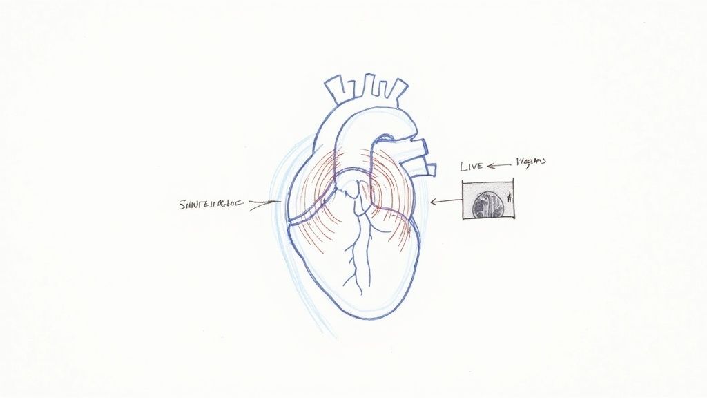
A heart echo scan, or to give it its proper name, an echocardiogram, is a safe and completely painless ultrasound test that gives us a live, moving picture of your heart. It uses sound waves to show exactly how your heart's chambers and valves are working, helping doctors diagnose a whole range of heart conditions without using any radiation.

If your doctor has suggested a heart echo scan, it’s a brilliant, proactive step towards getting a proper handle on your cardiovascular health. The best way to think of it is like a ship's sonar. The probe sends out harmless, high-frequency sound waves that bounce off your heart's structures, creating a detailed, real-time image on a monitor.
This scan lets your doctor see your heart in action, all without making a single incision. They can check its size and shape, and, most importantly, see how well it's pumping. It's an invaluable window into your heart’s function, and because it’s non-invasive and avoids X-rays, it's an incredibly safe and routine diagnostic tool.
A heart echo scan is one of the most vital diagnostic tools we have, often carried out as part of specialized radiology services or within dedicated cardiac imaging departments. Its main job is to help doctors spot and keep an eye on various heart conditions.
It offers some key insights:
Echocardiography is a cornerstone in managing cardiovascular diseases (CVD), which affect an estimated 7.6 million people in the UK. Tragically, CVD is responsible for around 64,000 premature deaths every year in the UK, making early and accurate diagnosis more critical than ever.
Ultimately, this scan gives your specialist the detailed information they need to protect your heart. Whether it's diagnosing a new problem or monitoring a known condition, it’s a foundational test in modern cardiology. The information we gain helps shape a precise and effective treatment plan that’s right for you.

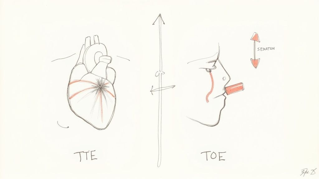
If your doctor has recommended a heart echo scan, you should know there isn't just one type. The two most common are the Transthoracic Echocardiogram (TTE) and the Transoesophageal Echocardiogram (TOE), and the right one for you depends entirely on what your specialist needs to see.
Think of it like taking a photograph. You can take a clear picture from across the room, or you can get right up close for the finer details. Both give you a picture, but they serve different purposes. Your cardiologist will choose the best angle to get the clearest view of your heart's health.
The Transthoracic Echo (TTE) is the go-to heart scan for most situations. It's the one most people are familiar with—completely non-invasive, painless, and straightforward.
During a TTE, a sonographer simply applies a cool gel to your chest and glides a small, handheld probe (a transducer) across the skin. This probe uses sound waves to create a live, moving image of your heart on the monitor. It’s an excellent way to get a solid overview of your heart's structure, see how the valves are working, and check its pumping strength.
The whole process is comfortable and requires no special preparation. It’s the same reliable technique we use for younger patients, which you can read about in our guide to a paediatric echocardiogram.
Sometimes, we need to get past obstacles like the ribs, lungs, or body tissue, which can slightly obscure the view from the outside. When a crystal-clear, detailed image is essential, the Transoesophageal Echo (TOE) is the answer.
With a TOE, a tiny transducer is fitted to the end of a thin, flexible tube. To ensure you're completely comfortable, your throat is numbed and you're given a light sedative to help you relax. The tube is then gently passed down your oesophagus (the pipe connecting your throat to your stomach).
Because your oesophagus sits directly behind your heart, this technique gives us an uninterrupted, high-definition view. It’s the best way to get a close-up look at the intricate workings of the heart valves or to check for tiny blood clots.
While it might sound a bit more involved, a TOE is a very common and safe procedure. The sedation keeps you relaxed, and the scan itself is usually over in 15-30 minutes. Your medical team will walk you through every step beforehand, making sure you feel at ease.
To help you see the differences at a glance, here’s a quick comparison of the two main types of scans.
This table provides a side-by-side comparison of the two main types of heart echo scans, helping you quickly understand the key differences in procedure, preparation, and purpose.
FeatureTransthoracic Echo (TTE)Transoesophageal Echo (TOE)ProcedureA transducer is moved across the chest.A small probe is guided down the oesophagus.InvasivenessNon-invasive and painless.Minimally invasive; requires light sedation.PreparationNone needed. Just wear loose clothing.Fasting for 4-6 hours beforehand.Image QualityGood general overview of the heart.Exceptionally clear and highly detailed.Best ForRoutine checks, assessing overall heart function.Detailed valve inspection, clot detection.DurationAbout 30-60 minutes.About 15-30 minutes for the scan itself.
Ultimately, the choice between a TTE and a TOE is always driven by one thing: getting the most accurate information needed to look after your health.
Knowing what to do before your appointment helps everything run smoothly and takes a lot of the stress out of the day. The steps are straightforward and change slightly depending on whether you’re having a standard transthoracic (TTE) or the more detailed transoesophageal (TOE) heart echo scan.
For a standard TTE, there’s almost nothing you need to worry about. You can eat, drink, and take your regular medications just as you normally would, unless your doctor has given you specific instructions to do otherwise. The only real practical tip is to wear comfortable, loose-fitting clothes, as you might be asked to change into a hospital gown.
Preparing for a TOE is a little different because it involves sedation. To make sure you’re safe and comfortable, there are a few extra steps to follow.
Here’s what you need to do:
Proper preparation is key to a successful scan. Following these simple guidelines helps our clinical team get the clearest possible images of your heart while ensuring you remain comfortable and safe throughout the process.
Feeling confident about your upcoming procedure starts with knowing what to expect. Getting the practical details sorted beforehand lets you focus on what really matters—your health. If you have any questions or need to check your appointment details, you can easily book appointments online at The Vesey or give our team a call.
When you step into the room for your heart echo scan, the first thing you’ll probably notice is that it’s calm and dimly lit. This isn’t to help you nod off, but to give the sonographer a crystal-clear view of the monitor, ensuring they capture the best possible images of your heart.
You'll be asked to lie down on an examination couch, usually on your left side. The sonographer, a specialist highly trained in ultrasound imaging, will then apply a small amount of clear, water-based gel to your chest. It might feel a bit cool at first, but it’s completely harmless and absolutely essential for the scan to work.
So, what’s the gel for? Its main job is to get rid of any tiny air pockets between your skin and the ultrasound probe, which is called a transducer. This is crucial because it ensures the sound waves can travel smoothly into your body and back again, which is how we get a sharp, detailed picture of your heart.
Once the gel is on, the sonographer will gently place the transducer on your chest. They’ll move it around, applying light pressure to see your heart from different angles—a bit like a photographer finding the perfect position for a portrait. They are methodically capturing images of your heart's chambers, valves, and major blood vessels to build a complete picture of its health.
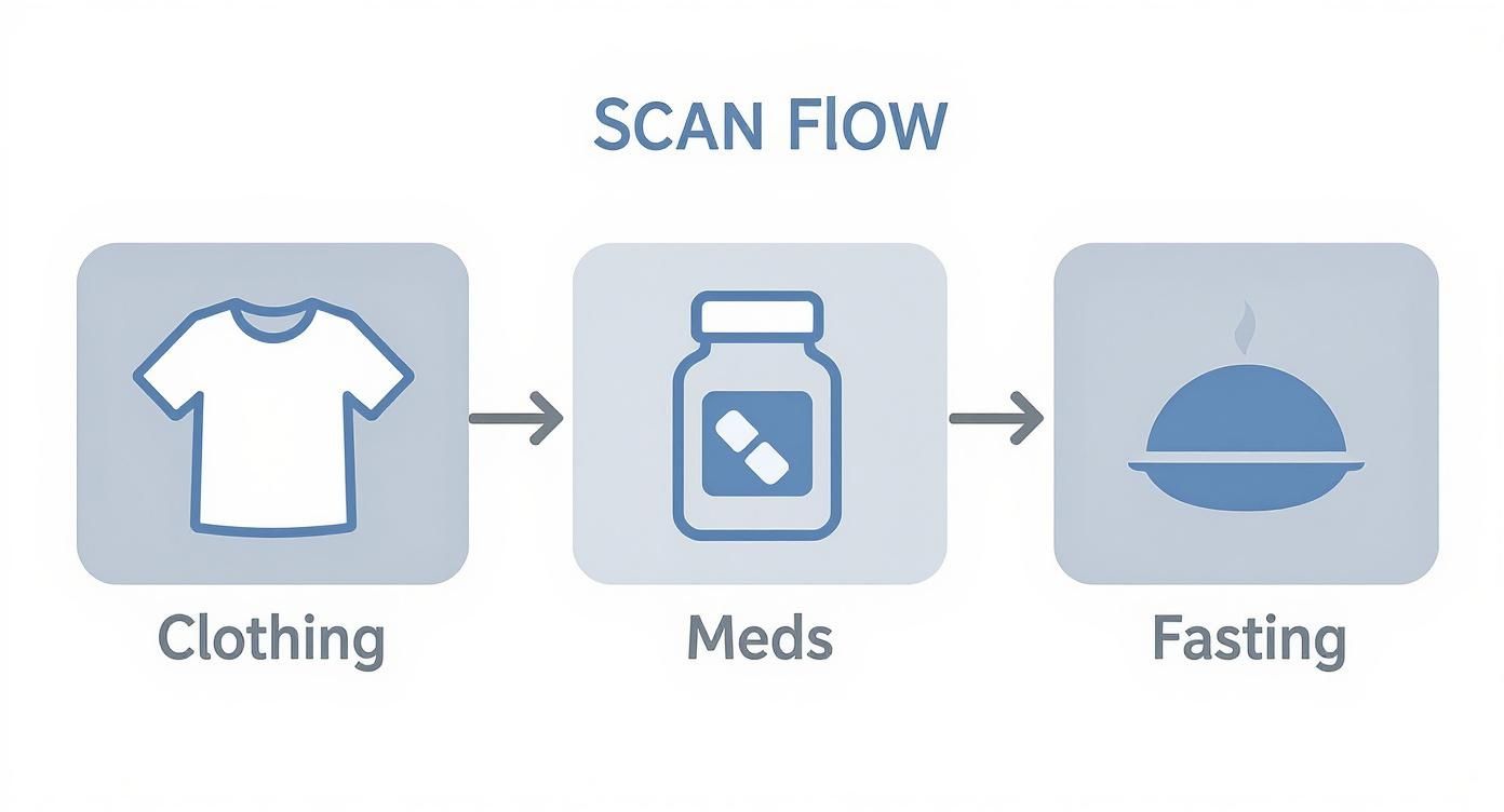
This visual guide shows the simple, key steps often involved in preparing for a scan, from clothing choices to fasting instructions for specific procedures.

As the infographic shows, the preparation is usually quite straightforward and just depends on the exact type of echo scan you’re having.
As the sonographer moves the probe, you’ll probably hear a rhythmic ‘swooshing’ sound coming from the machine. Don't worry, this is completely normal. In fact, it's a key part of the Doppler ultrasound technology being used.
That sound is actually your blood flowing through the heart's chambers and valves. It's not just noise; it’s providing real-time data, allowing the sonographer to:
So while you’re lying there, that swooshing noise is giving the team a huge amount of information about your heart’s performance. The whole thing is quite interactive. The sonographer might ask you to hold your breath for a few seconds or shift your position slightly. This just helps them get a clearer view by moving things like your lungs or ribs out of the way.
A standard transthoracic echocardiogram is typically done and dusted within 30 to 60 minutes. It's a completely painless and safe procedure, designed to be as comfortable as possible while gathering all that vital information.
By understanding each step—from the dim lighting to the cool gel and the meaning behind the sounds—the entire process becomes a lot less mysterious. It's a straightforward, non-invasive way for us to get a comprehensive look at your heart, and the procedure is designed to be as stress-free as possible for you.
Once your heart echo scan is done, the next step is looking at the report. Getting a document full of medical jargon can feel a bit overwhelming, but it’s best to think of it as a detailed story about your heart's health. Understanding its key chapters can empower you for the conversation with your cardiologist.
Think of the report as a performance review for your heart. It doesn't just give a simple "pass" or "fail"; it breaks down specific metrics that show exactly how well each part is doing its job. Your doctor will translate these numbers into a meaningful plan, but let’s get familiar with the main characters in this story.
One of the most important numbers you'll come across is the Ejection Fraction (EF). This is a percentage that measures your heart's pumping power. If you imagine your heart as a squeezy bottle, the Ejection Fraction tells you what percentage of blood is pushed out with each contraction.
A typical, healthy heart has an EF between 55% and 70%. This doesn’t mean your heart is only working at half-steam—it's perfectly normal for a healthy amount of blood to stay in the ventricle after each beat. A lower EF, however, might suggest the heart muscle isn't pumping as strongly as it should, which is a key indicator for conditions like heart failure.
Another major focus is your heart valves. The report will describe their structure and how they're working, noting if they open and close properly. You might see terms like "stenosis" (a valve is too narrow and stiff) or "regurgitation" (a valve is leaky), which are used to flag any issues.
Your report is much more than just a list of data points. It’s a structured assessment that helps your doctor make informed decisions, turning images and sounds into actionable insights about your cardiovascular health.
Finally, the report details the size and thickness of your heart’s chambers. An enlarged chamber or a thickened heart wall can be a sign that the heart has been working under strain, maybe because of high blood pressure or a faulty valve.
The findings in your report are standardised to make sure they are clear and consistent. In the UK, reports often follow guidelines from the British Society of Echocardiography, detailing key parameters like left ventricular ejection fraction, valve function, and chamber sizes. These findings are crucial because they directly guide what happens next—from taking no action at all to starting medication or being referred for more tests. You can discover more about how these reports shape patient treatment plans.
It's important to remember that a heart echo scan is just one piece of the puzzle. Your cardiologist will combine these results with other information, like your symptoms, a physical examination, and potentially other diagnostic tests. For a broader look at cardiovascular diagnostics, you might be interested in our guide to comprehensive heart health blood tests.
Your results are the beginning of a conversation, not the final word. They provide the vital information your specialist needs to build a complete picture of your health and work with you on the best path forward.
It’s completely understandable to ask about the safety of any medical procedure, even one as common as a heart echo scan. The good news is that a standard transthoracic echocardiogram (TTE) is one of the safest diagnostic tools we have in modern medicine.
The scan is entirely non-invasive. It uses the exact same ultrasound technology that’s been used for decades to safely monitor babies in the womb. Because it works by sending harmless sound waves into the body to create pictures, there is zero radiation exposure. For a standard TTE, there are no known risks or side effects, making it an incredibly reliable way to get a clear look at your heart.
The more detailed transoesophageal echo (TOE) is also considered a very low-risk procedure, especially as it’s always carried out by a highly trained specialist. Because a small probe is gently guided down your food pipe (oesophagus), there are a couple of very minor potential risks, but these are rare.
The most common thing people experience is a mild, temporary sore throat, which almost always clears up within a day or two. There's also a tiny chance of a minor scratch to the lining of the throat.
It's important to see these minimal risks in the context of the significant diagnostic benefits. A TOE gives us an exceptionally clear, unobstructed view of the heart's structures, which is often crucial for accurately diagnosing complex valve problems or finding small blood clots that a standard echo might miss.
Your medical team will talk you through everything beforehand, answering any questions you have and taking every possible precaution to keep you safe and comfortable. The safety record for both types of heart echo scan is outstanding, and the information they give us is vital for protecting and managing your long-term heart health.
It's completely normal to have a few questions before any medical procedure. To help you feel more comfortable and prepared, we’ve put together answers to some of the most common queries we hear from patients about their heart echo scan.
A standard transthoracic echo is a pretty quick and straightforward process. All in, you can expect it to take between 30 and 60 minutes. The more detailed transoesophageal echo (TOE) might be a bit faster for the scan itself, but it does need extra time for preparation and for you to recover from the light sedation afterwards.
One of the biggest worries people have is whether the scan will be painful. Let me put your mind at ease: a standard heart echo scan is completely painless. The only things you’ll feel are the cool gel on your skin and the gentle pressure of the probe moving across your chest. For a TOE, we numb your throat and use sedation to make sure you're comfortable the entire time.
Once the scan is done, a specialist cardiologist will carefully analyse the images. Your consultant will then walk you through the full report at your follow-up appointment, which is usually scheduled within a week or two. This gives them enough time to build a complete picture of your heart's health.
It's also quite common to need a repeat echo scan down the line. This isn't usually a cause for alarm. It could be to monitor a known condition, check how well a specific treatment is working, or simply to get a clearer picture if the first images weren't quite perfect. It’s a routine part of managing long-term heart health. For a deeper dive into proactive cardiac care, our guide on what's involved in a private heart check is a great resource.
The demand for diagnostic imaging is always on the rise. To give you an idea, in England, diagnostic ultrasound—which includes echocardiography—accounted for around 0.89 million tests in April 2025 alone. This really highlights its vital role in modern healthcare. You can learn more about these NHS diagnostic imaging statistics.
At The Vesey, our dedicated cardiology team is here to support you every step of the way. If you have any further questions or wish to book a consultation, please visit us at https://www.thevesey.co.uk.

At The Vesey, our dedicated cardiology team is here to support you every step of the way. If you have any further questions or wish to book a consultation, please visit us at https://www.thevesey.co.uk.
