.webp)
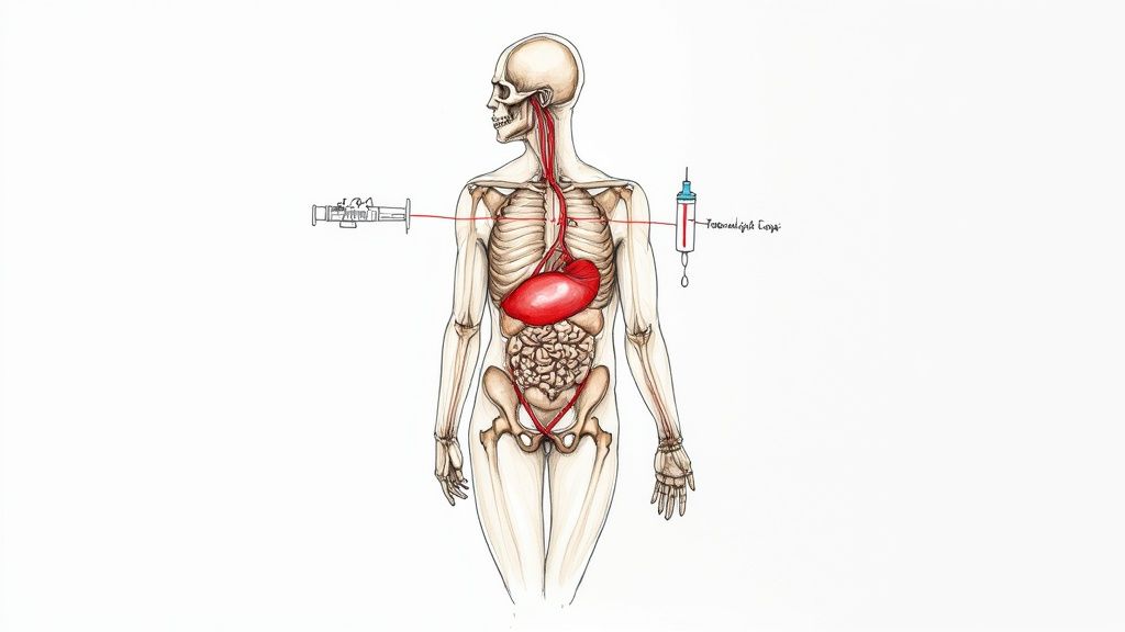
An MRI scan with contrast isn't just a standard imaging procedure; it’s a high-definition upgrade that uses a special dye to make certain parts of your body pop with detail. This extra clarity gives radiologists a much sharper, more defined picture, which is often the key to getting the diagnosis right.

Think of a regular MRI as a beautifully detailed black-and-white photograph of your body’s inner workings. It's incredibly useful, but sometimes, different tissues can look quite similar, making it tough to spot an abnormality hiding in plain sight. An MRI scan with contrast is like giving the radiologist a set of highlighters.
We introduce a special substance—a contrast agent, usually containing a rare earth metal called gadolinium—and certain tissues absorb it, making them "light up" on the scan images. Suddenly, those areas stand out with brilliant clarity against everything else. The black-and-white photo now has specific, vivid highlights pointing exactly where we need to look.
This enhanced visibility isn’t just about making the pictures look clearer; it’s about uncovering crucial information that might otherwise stay hidden. The contrast agent is given through a small intravenous (IV) line and travels through your bloodstream. It has a knack for accumulating in areas with increased blood flow or where the normal barriers between blood vessels and tissues have broken down.
This makes it especially good at pinpointing:
A contrast-enhanced MRI doesn't just show us anatomy. It gives us a functional map, revealing not just what is there, but how active it is by tracking where blood flow is most dynamic. That distinction is often the key to a definitive diagnosis.
The precision you get with MRI scans with contrast makes them an essential tool in the UK's healthcare system, particularly in fields like neurology, oncology, and cardiology. When investigating a potential brain tumour, for example, the contrast helps define its exact size, shape, and how it sits next to healthy brain tissue. Our detailed guide on what a brain MRI scan shows dives deeper into this.
The diagnostic power is clear from how often these scans are performed. In England, MRI is a cornerstone of medical imaging, with NHS data showing 380,000 tests were carried out in April 2025 alone. A huge portion of these rely on contrast agents to get the necessary detail, especially in urgent cancer pathways where every bit of precision counts. This is why your doctor might specifically request this more advanced type of scan to get the clear answers needed for your care.
If your doctor has specifically requested an MRI scan with contrast, it’s because they need to see more than just a static picture of an organ or tissue. They need to understand what it’s doing.
The contrast agent, which usually contains a substance called gadolinium, acts like a dynamic highlighter. Once it enters your bloodstream via an IV, it naturally collects in areas with unusual activity—like increased blood flow or a breakdown in the normal barriers between blood vessels and tissues. This is what lets it reveal processes a standard scan would completely miss.
Think of it like this: a standard MRI gives you a detailed blueprint of a building. Adding contrast is like switching on the power to see which rooms are lit up, where the electricity is flowing, and which circuits might be faulty. It transforms a static map into a live feed of what’s happening inside your body.
This ability to highlight active areas is what makes contrast-enhanced MRI so crucial for diagnosing and monitoring a huge range of conditions. It gives your medical team the clarity they need to make confident decisions about your health and guide treatment with real precision.
Some key real-world applications include:
This level of diagnostic detail is central to modern medicine. The UK market for MRI contrast media is a key part of a global industry estimated to be worth around USD 1.66 billion in 2025. It’s expected to grow by over 6% each year, driven by the increasing need for detailed diagnostics. You can learn more from the full market report on MRI contrast media.
To make this clearer, here’s a quick breakdown of why contrast might be used for different parts of the body.
This table isn’t exhaustive, but it gives you a good idea of how a contrast agent provides targeted information that is critical for an accurate diagnosis.
Beyond organs and soft tissues, contrast agents are exceptionally good at mapping out your circulatory system. This specialised scan is called a Magnetic Resonance Angiography (MRA). The contrast agent essentially lights up your arteries and veins, allowing radiologists to create a detailed road map of your blood vessels.
This isn't just a simple picture; it's a functional assessment. A contrast-enhanced MRA can reveal narrowing, blockages, or aneurysms (bulges in vessel walls) with remarkable accuracy, often eliminating the need for more invasive procedures.
It's particularly useful for assessing conditions like peripheral artery disease, where blood flow to the limbs is restricted. Understanding the precise location and severity of a blockage is the first step towards getting the right treatment. If you'd like to understand more about this, you can read our guide on the diagnosis and treatment of arterial disease.
By connecting the procedure directly to your health concerns, it becomes clear why MRI scans with contrast are so frequently recommended. They provide your medical team with the specific, detailed information needed to move from uncertainty to a clear and actionable diagnosis.
Knowing what to expect before any medical procedure can make a world of difference, and an MRI with contrast is no exception. A bit of preparation helps everything run smoothly and can really set your mind at ease.
Think of it this way: you’re an active partner in your own healthcare. This guide will walk you through the simple, practical steps to get ready, making sure your appointment is as safe and effective as possible.
The first questions are usually about food and clothing. For most MRI scans with contrast, you can eat and drink normally beforehand. Simple. However, if your scan is focused on the abdomen or pelvis, you might be asked to fast for a few hours. Your appointment letter will have all the specific instructions, so always give that a careful read.
When it comes to what you wear, the golden rule is no metal. The MRI machine is essentially a giant, powerful magnet.
To take the guesswork out of it, most clinics will simply give you a hospital gown to change into. This guarantees you're completely safe to enter the scanner.
This is, without a doubt, the most important part of your preparation. Because of the strong magnetic field and the contrast dye, the radiology team needs a crystal-clear picture of your medical history. Being open and thorough here is vital for your safety.
You’ll be given a detailed safety questionnaire. It’s essential you fill this out honestly and completely—even small details you think might not be relevant can be important.
Your medical history is the key to a safe scan. Every detail you provide, from past surgeries to current medications, helps the team tailor the procedure specifically to you, ensuring the highest standards of safety and care.
Be ready to chat with the radiographer about a few key things:
By running through these steps, you’re not just a patient waiting for a scan; you're an informed and crucial part of the process. For more general details on what to expect during your visit, have a look at our comprehensive patient information guide.
Knowing what happens during an MRI scan with contrast can make the whole experience feel much less daunting. So, let’s walk through the process step-by-step, from the moment you arrive to the moment you’re done. This way, you’ll feel confident and fully prepared.
When you get to the clinic, you’ll check in at reception and might have some final paperwork to sort out. Soon after, a radiographer—the specialist who operates the MRI scanner—will come and greet you. They’ll run through your safety questionnaire one last time, double-checking your medical history and answering any questions you might have. It's a final, vital check to ensure everything is safe.
This quick infographic sums up the main things to keep in mind before you arrive.

As you can see, thinking about what you eat, wearing metal-free clothes, and being open about your health history are the keys to a smooth appointment.
Once the safety checks are out of the way, it’s time for the contrast agent. This is given through a small intravenous (IV) line, often called a cannula. A tiny, flexible tube is inserted into a vein, usually in your arm or the back of your hand. You’ll feel a quick scratch, much like having a blood test.
With the line in place, the contrast agent is injected. It’s very common to feel a temporary cool or cold sensation moving up your arm as the fluid goes in. Some people also notice a brief metallic taste or a feeling of warmth that spreads through their body. These are all completely normal and expected sensations that disappear in a moment or two.
The administration of the contrast agent is a straightforward and quick part of the process. The sensations are temporary and expected, so being aware of them beforehand can help you feel more relaxed and in control.
After the contrast agent is in, the radiographer will lead you to the MRI machine. It’s a large, tunnel-like scanner with a flat, motorised bed that slides into the centre. They’ll help you get comfortable on the bed, using pillows or cushions to make sure you’re in the perfect position and can stay there.
When you’re settled, the bed will slowly move into the scanner. The radiographer will be in the next room, but you’re never truly alone—you can talk to them through an intercom the entire time, and they’ll be watching you through a large window.
The first thing you’ll probably notice is the noise. The machine makes a series of loud, repetitive knocking, buzzing, and whirring sounds while it works. This is just the sound of the scanner capturing the images. To make it more comfortable, you'll be given earplugs or noise-cancelling headphones, and you can often listen to music to help pass the time.
Staying as still as possible is crucial for getting clear, high-quality images. Even tiny movements can blur the pictures, which might mean having to repeat parts of the scan. An MRI can take a little while, similar to other types of imaging like a CT scan, so having a few tricks up your sleeve to stay relaxed is a great idea. If you're interested, you can find out more about how long different scans can take in our other article.
Here are a few proven techniques to help you stay calm and still:
By understanding what each step involves and having a few coping strategies ready, you can go into your MRI scan with contrast feeling confident and knowing exactly what’s ahead.

It’s completely normal to have questions about the safety of any medical procedure, especially when it involves an injection. When it comes to MRI scans with contrast, the key takeaway is that for the vast majority of people, the procedure is remarkably safe. The contrast agents we use today have been refined over decades to maximise diagnostic clarity while keeping risks to an absolute minimum.
The whole process is designed around your wellbeing, starting with that detailed screening questionnaire you fill out beforehand. This isn't just paperwork; it’s a critical tool that gives the radiology team a clear picture of your health, making sure the scan is right for you. Your honest input here is a cornerstone of the safety protocol. For a deeper look at this, our guide on the importance of informed consent in healthcare is a great resource.
Most people who receive a gadolinium-based contrast agent don't experience any side effects at all. If reactions do happen, they are almost always very mild and disappear quickly on their own.
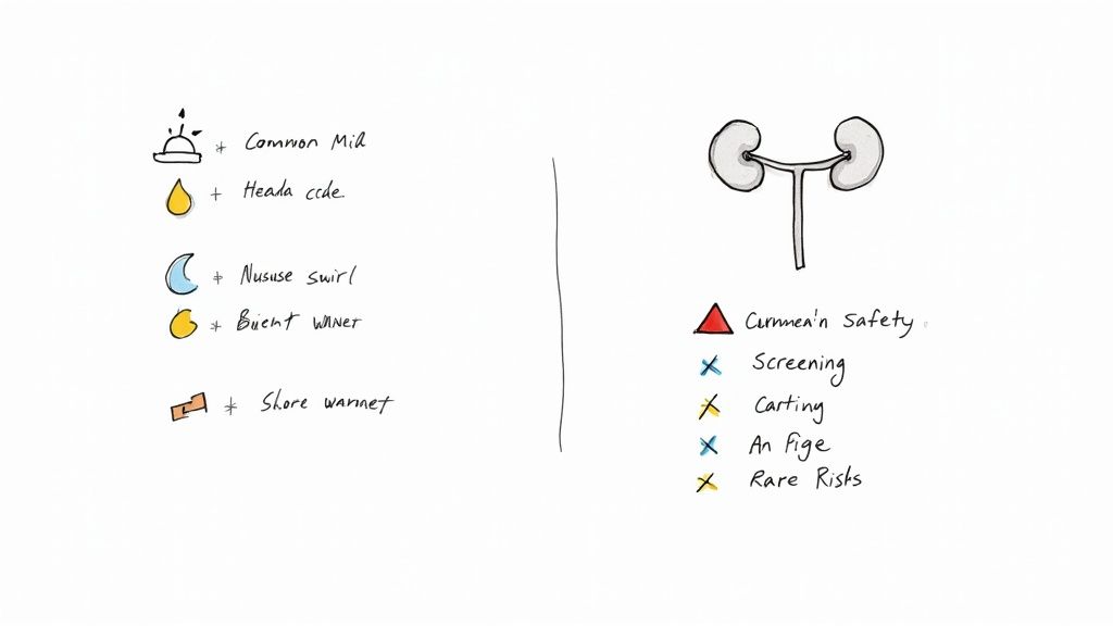
Think of them as brief, minor inconveniences rather than genuine problems. The most common sensations include:
These sensations usually fade within an hour or two after your scan. Drinking plenty of water afterwards helps your body flush the agent out and can ease any mild symptoms even faster.
While serious side effects are incredibly rare, it’s still important to be aware of them. The medical team is fully trained and equipped to handle any situation that might pop up.
Severe allergic reactions (anaphylaxis) are exceedingly uncommon but possible. Symptoms might include breathing difficulties, swelling of the face or throat, or a sudden drop in blood pressure. This is precisely why you're monitored so closely by trained professionals during and right after the injection.
Patient safety is the absolute top priority. Radiology departments are prepared emergency environments. The on-site team has the expertise and equipment to manage any adverse reaction immediately, ensuring your wellbeing from start to finish.
To put the risks into perspective, here's a quick comparison of potential reactions.
This table shows that while a spectrum of reactions is possible, the ones that cause real concern are exceptionally rare and the medical team is fully prepared to manage them.
You may have come across information about a condition called Nephrogenic Systemic Fibrosis (NSF). This is a serious but now exceptionally rare disease that causes thickening of the skin and connective tissues. It was historically linked to older types of gadolinium agents, specifically in patients with severe, pre-existing kidney disease.
Modern screening protocols and improved contrast agents have virtually eliminated this risk in the UK. We always perform a thorough check of your kidney function before giving any contrast if there's any reason for concern. This careful, evidence-based approach is a perfect example of how medical practice evolves to protect patients.
In recent years, research has shown that tiny amounts of gadolinium can remain in the body, including the brain, long after an MRI scan with contrast. This is often referred to as gadolinium retention.
Right now, there is no solid evidence suggesting these trace deposits cause any harm in patients with normal kidney function. Medical bodies across the globe, including here in the UK, continue to monitor all new research. The consensus remains that the diagnostic benefit of a necessary contrast-enhanced MRI far outweighs the theoretical risks of retention. To minimise any potential issues, clinicians always use the lowest effective dose and only recommend contrast when it is truly essential for an accurate diagnosis.
Medical imaging is far from static. The technology behind MRI scans with contrast is moving forward at a blistering pace, with researchers hunting for ways to make scans faster, images sharper, and the whole experience safer and more comfortable for patients. These developments give us a glimpse into a future where diagnostics are more precise than ever.
One of the most exciting areas of research is in the contrast agents themselves. Scientists are developing next-generation dyes that are far more stable and targeted. Think of them as "smarter" agents, designed to deliver exceptional image clarity while being flushed out of the body even more efficiently. This would further minimise the already tiny risks tied to today's agents.
But beyond the chemistry, the biggest waves are being made by artificial intelligence (AI) and deep learning. These aren’t just buzzwords; they’re powerful tools poised to completely reshape the patient journey.
What if you could get the same high-quality diagnostic images from your MRI, but in half the time? That’s the core promise of AI in medical imaging. We all know that long scan times can be tough. They increase the chances of tiny movements blurring the images and add to patient anxiety and discomfort.
AI algorithms are now being trained to build a crystal-clear picture from far less raw data. A scan that once took 45 minutes could soon be done in 15 or 20, without sacrificing a single ounce of diagnostic detail.
This speed-up has huge implications, especially for the UK's healthcare system. Shorter scan times could ease the burden on busy NHS and private clinics, helping to cut down waiting lists and get more people the crucial diagnostics they need, sooner.
A great example is the latest work in multi-contrast MRI, which captures several image types in a single, long session. A new deep-learning method is showing real promise in speeding up these scans, which are vital for diagnosing complex conditions like Alzheimer's disease.
The integration of AI aims to solve a fundamental challenge: achieving maximum image quality in minimum time. For patients, this translates directly into a more comfortable experience and faster access to care.
These technological leaps are also transforming how medical information is handled behind the scenes. It's not just about the scanner; the future also involves big improvements in processes like healthcare documentation automation, which will make managing scan results and patient records far more seamless.
When you combine faster scans, better contrast agents, and smarter data management, you get a vision of healthcare that’s more responsive and personalised. For patients, this all points to one thing: quicker diagnoses, more effective treatment plans, and a much less stressful path through the diagnostic process.
It’s completely normal to have questions before any medical procedure. We’ve put together clear, straightforward answers to some of the most common queries we hear from patients about having an MRI with a contrast agent.
No, the scan itself is entirely painless. The only part you might feel is the very brief, small scratch from the IV needle when the contrast dye is introduced, much like having a blood test.
Some people mention a temporary cool or even warm flushing feeling as the agent enters the bloodstream. This is perfectly normal, doesn't hurt at all, and passes in just a few moments. For most patients, the main challenges are simply dealing with the loud noises the machine makes and the need to lie perfectly still.
For anyone with healthy kidneys, the body is very efficient at clearing the gadolinium-based contrast agent. It's filtered out of your system naturally through your urine, with the vast majority gone within 24 hours.
Your clinical team will almost certainly encourage you to drink plenty of water after your scan. This simply helps your kidneys flush the agent out of your system a bit faster. The pre-scan screening is so important because it identifies anyone with kidney issues, as they would process and clear the agent more slowly.
Yes, you can. The current guidelines from the Royal College of Radiologists state that it is safe to continue breastfeeding after receiving a gadolinium-based contrast agent.
An extremely small amount of the agent may enter the breast milk, and an even smaller fraction of that is absorbed by the baby. This trace amount is not considered harmful. You should always confirm the most current advice with your doctor and the radiology department.
Allergic reactions are rare, but the medical team is always fully prepared to handle them. Mild reactions, like some itching or hives, are the most common and are easily treated with simple antihistamines.
Severe reactions are extremely rare. Rest assured, the on-site clinical team is trained and has emergency medications ready to manage any situation immediately. You are monitored closely during and after the injection to ensure your complete safety.
At The Vesey, our priority is making sure you feel informed, comfortable, and cared for at every stage. If you have more questions or want to arrange a consultation for rapid access to diagnostic imaging, please visit us at https://www.thevesey.co.uk.

